

We use the most up-to-date technology to ensure the best eye care possible. Here are some of the different types of tests and equipment you may experience on a visit to our Practice.

Epic 5100
This sophisticated, very streamlined, electronic refraction system allows us to refract for glasses and contact lens prescriptions much faster and more easily for you than is possible with conventional, manual refracting equipment. At the push of a button, the refracting tech can show you your new glasses prescription compared to your old glasses prescription. What people like the best is that instead of having to remember what the 2 different image choices (“1 or 2”) looked like, they are presented simultaneously side by side. You can study them and choose one or the other. It is a much faster, much easier way to test for your glasses prescription.
OPD 3 Wavefront Aberrometer
This instrument can make a number of important measurements of the optics of your eyes almost instantly. It gives us a very close estimate of your glasses prescription, measures the curvature of your cornea-a very important optical surface of your eye, measures the optical characteristics of the lens inside your eye, and can detect and measure defects in the optics of the eye we didn’t even know existed before this technology was developed. This gives us much more information about the optics of your eye than was possible in the past.


Cirrus 6000 OCT
One of the greatest breakthroughs in ocular diagnostic technology, this amazing instrument can take highly magnified cross-section images of any part of the eye’s structure- the cornea and anterior chamber, any part of the retina, and especially the optic nerve and the macula, without touching the patient. It has had a huge impact on our ability to diagnose, treat, and monitor glaucoma, macular degeneration, diabetic retinopathy, optic nerve diseases, and MS. We use it extensively in telemedicine, where we email the images to sub-specialists for a virtual consult, often saving the patient an unnecessary trip, and allows us to manage many cases ourselves that we once would have had to refer out.
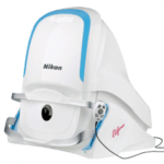
Optos California
Another amazing breakthrough in diagnostic imaging technology, the Optos can take wide-field images of nearly the entire retina all at one time through normal-sized pupils- it is often not necessary to dilate the pupils in order to get a good image of the peripheral retina, which was impossible prior to the development of this technology. It is very important to rule out the presence of asymptomatic retinal breaks, tears, hemorrhages, and tumors in the retinal periphery, which always required widely dilated pupils in the past. It enables us to take better care of you, and saves a significant amount of your time, too. Like the OCT images, we often use these Optos images for telemedicine with sub-specialists, again often saving the patient a trip, and often allowing us to manage the patient locally. We store these images so that we can compare them from one year to the next, monitoring for changes that could indicate problems.

HFA 3 Visual Field Tester
This instrument performs a very important test called a threshold visual field. It tests the central 25 degrees of the patient’s field of vision, searching for evidence of very subtle vision changes or loss in sensitivity that the patient is almost never aware of. It helps us tremendously in diagnosing diseases like glaucoma, neurological diseases, and often will find the evidence of the presence of a brain tumor or stroke, usually while the disease process is still completely asymptomatic. It is a very powerful tool that allows us to take even better care of your eye health and your vision
Slit Lamp Biomicroscope
The most important diagnostic instrument of all for eye doctors is undoubtedly the slit lamp biomicroscope. It allows us to see the structures of the eye under extremely high magnification. It is a powerful and indispensable diagnostic tool for determining whether the eyes are normal and healthy, and especially for determining what the underlying cause is of an eye infection, inflammation, foreign body sensation, or eye injury, all of which are very difficult or impossible to determine without the high magnification and illumination the slit lamp provides the doctor.


Handheld Slit Lamp Biomicroscope
Unfortunately, if the patient is unable to physically get their chin and forehead onto the chin rest and forehead rest of the standard instrument stand slit lamp (which is often the case with wheelchair patients), our ability to properly diagnose and care for them is severely compromised. Fortunately, we have and use a portable, handheld slit lamp precisely for these situations, which allows us to properly care for those with significant physical limitations. And we are of course wheelchair friendly and accessible throughout our office.
Digital Retina Imaging
We take images of the retina, macula, and optic nerve of all our patients age 10 and over. This is not considered the standard of care in the industry, but we consider it OUR standard of care, and it is priceless for monitoring the health of the retina, macula, and optic nerve. As they say, a picture is worth a thousand words!


Handheld Retinal Camera
Unfortunately, we often encounter the same problem with wheelchair patients that we do with the slit lamp- they can’t physically get into the instrument for such imaging. However, we do have a handheld retina camera that allows us to take retinal images of these patients despite their physical limitations.
The Handy-Ref Handheld Autorefractor/Auto Keratomer
Again, patients who are physically unable to reach the chin rest and forehead rest of the OPD3 Autorefractor mentioned above can be effectively autorefracted with our handheld Handy-Ref autorefractor. Wheelchair patients can be examined without even having to be transferred from their wheelchairs.


Meibox Meibographer
This allows us to diagnose ocular surface disease (formerly referred to as “dry eye”) much more accurately and much sooner in the process. The crux of the problem is malfunctioning Meibomian glands (oil glands) in the eyelids. Healthy Meibomian glands are crucial for ocular health, it was very poorly understood until very recently. Treatment of Meibomian gland dysfunction has been a breakthrough in the treatment and management of the ocular surface disease.
Forum Integrative Network Software
We have a very sophisticated network diagnostic software that can take the results of our various imaging instruments for a given patient and integrate and analyze the data. This augments our ability to diagnose eye diseases earlier in the process and allows us to take even better care of you than we could before.
